
5
Strategies for Monitoring Cognitive Performance
Up to this point the central focus of this report has been on the assessment or monitoring of the combat service member’s capacity to perform physical tasks. In this regard, the importance of factors that influence bone and muscle health, as well as other processes that underlie and optimize physical endurance and resistance to physical injury, have been highlighted, and for good reason. Clearly it is necessary to ensure that operational personnel are as physically fit as possible because success on the battlefield is to a great extent dependent on the ability of combat service members to carry and operate weapons, overcome physical obstacles, traverse distances in harsh environments, and endure a host of physical stresses and strains that could easily overwhelm unfit individuals. However, optimal performance in today’s military also is increasingly dependent on a high level of cognitive fitness. The widespread use of computerized weapon systems; complicated communications and targeting devices; high-performance aircraft, tanks, and maritime vessels; and the technologically advanced diagnostic systems used in the maintenance of military equipment demands the highest levels of cognitive readiness.
In the following sections, operator cognitive fatigue, one of the principal threats to military readiness, is discussed. Also included is an overview of the primary operational causes of fatigue, followed by a brief synopsis of strategies that should be considered for monitoring the cognitive status of servicemembers.
The fact that the focus here is on the fatigue that results from sleep deprivation should in no way imply that this is the only stressor of concern in the operational environment. As noted earlier in this report, combat service members are routinely exposed to a wide variety of physical and environmental stresses that, if ignored, will ultimately degrade operational performance. Heat stress and dehydration pose major threats to the cognitive readiness of ground combat service members, and these factors can be expected to exacerbate the fatigue from sleep loss and strenuous work. In the aviation arena, uncomfortable levels of noise, heat, vibration, and mental workload must be dealt with by pilots on a day-to-day basis, and these stresses likewise can be expected to compromise cognitive
capacity. However, since a detailed discussion of each of these areas is beyond the scope of this report, it is hoped that the reader can generalize many of the concepts from the forthcoming discussion of the most common cause of operator fatigue (sleep deprivation) to the fatigue stemming from other operational stressors.
THE PROBLEM OF SLEEPINESS AND COGNITIVE DEGRADATION IN MILITARY SETTINGS
Current military doctrine requires that units operate around the clock during times of conflict because the success of battlefield operations depends, at least in part, on maintaining the momentum of continuous day-night operations (U.S. Army, 1997). Technological advances, such as night vision devices, have enhanced the night-fighting capabilities of both ground and air combat military personnel, making around-the-clock missions a highly feasible component of the modern military strategy. Combining efficient day and night fighting capabilities across successive 24-hour periods places a significant strain on enemy resources and presents a clear tactical advantage for U.S. forces. In fact, the Air Force Chief of Staff recently noted that persistent and sustained operations “24 hours a day, seven days a week” are essential to attaining U.S. victory in today’s battle space (Elliott, 2001).
However, there are difficulties inherent in maintaining effective around-the-clock operations. For example, aircraft can function for extended periods without adverse effects, but human operators need periodic sleep for the restoration of both body and cognitive function (Home, 1978). Depriving humans of proper restorative sleep produces attention lapses and slower reaction times, which are associated with poor performance (Krueger, 1991). It has been determined that sleep-deprived personnel lose approximately 25 percent of their ability to perform useful mental work with each 24-hour period of sleep loss (Belenky et al., 1994). Thus by the end of 3 days without sleep, combat service members may be considered totally ineffective in the operational setting, especially if they are performing complex tasks, such as operating computerized command-and-control centers or flying an aircraft. This is a significant problem given that an Army manual makes it clear that “Soldiers in continuous operations can expect to be deprived of extended regular sleep, possibly any sleep, for as long as three to five days” (U.S. Army, 1991, P. 3–10).
Over the past several years the problem of sleep loss and fatigue has escalated because of increased requirements on military forces due to reductions in manpower and other resources. Over the past 10 to 15 years Army funding has been cut 38 percent and the number of personnel has been cut 35 percent, while missions have increased 300 percent (U.S. Army, 1996). A similar problem exists in the Air Force, where there has been a 37.7 percent reduction in military personnel and about a 50 percent reduction in the number of active Air Force tactical wings (Daggett and Belasco, 2002), while the operational tempo has
increased by as much as 400 percent (Correll, 1998). U.S. military capabilities are increasingly strained as understaffed units strive to accomplish more work with fewer resources. The ultimate result has been diminished military combat readiness (Spencer, 2000), in part because of increased levels of physical and cognitive fatigue.
Although reductions in available resources do not guarantee that sleep deprivation will be a problem in the operational environment, they create a situation in which the available personnel are more likely to work prolonged shifts without the benefit of sufficient rest. Krueger (1991) reported that the efficiency of combatants in sustained operations can be significantly compromised by inadequate sleep. Vigilance and attention suffer, reaction time is impaired, mood declines, and some personnel begin to experience perceptual disturbances. Naitoh and Kelly (1992) warned that poor sleep management in extended operations quickly leads to motivational decrements, impaired attention, short-term memory loss, carelessness, reduced physical endurance, degraded verbal communication skills, and impaired judgment. Angus and Heslegrave (1985) noted that cognitive abilities suffer 30 percent reductions after only 1 night without sleep, and 60 percent reductions after a second night.
Although all types of performance are not affected to the same degree by sleep loss, the fatigue from prolonged duty periods clearly is a threat to unit readiness in the operational context. This is especially the case for tasks that are lengthy, devoid of performance feedback, and boring. Caldwell and Ramspott (1998), Wilkinson (1969), and Wilkinson and colleagues (1966) found that when task durations extend beyond 15 to 20 minutes, performance deteriorations from fatigue become far more pronounced than when the task durations are shorter. Wilkinson (1961) found that knowledge of results alone can significantly attenuate the effects of sleep deprivation on some types of vigilance tasks. In addition, Wilkinson (1964) reported that while reaction-time tasks and vigilance tasks are most degraded by sleep loss, more interesting learning tasks and performance tasks often are less affected, presumably because the subject’s level of interest provides greater motivation and ability to resist attention lapses or outright sleep episodes (although, as warned by Dinges and Kribbs [1991], this works only up to a point).
In addition to the impact of task characteristics, it should be noted that there are individual differences in resiliency to sleep loss. Although this is an area that has not been sufficiently researched at this point, Van Dongen and colleagues (2003) found that there are basic interindividual differences in vulnerability to sleep debt that cannot be explained on the basis of differences in sleep need (i.e., short vs. long sleepers). This source of variability no doubt contributes to findings that there are wide differences in the accuracy with which several currently available methods can predict performance decrements. As the reader later reviews the strategies proposed to monitor alertness in the field, a recent U.S. Highway Traffic Safety report should be kept in mind (Dinges et al., 1998). In that report, even the eye-closure measure PERCLOS, which was found to be one
of the most predictive indicators of fatigue-related performance lapses under laboratory conditions (r=0.7), sometimes correlated with performance only at about r=0.3 in some individuals. Two electroencephalographic (EEG) algorithms showed a median predictive capability of only 0.3 to 0.4, and one type of eye-blink monitoring device correlated with minute-to-minute lapse frequency at a median level of 0.17. Thus despite the known dangers of fatigue and the established need to accurately measure it in some contexts, it is clear that much work remains to be done on monitoring technologies that can accurately predict moment-to-moment performance fluctuations. Clearly sleep loss from prolonged duty periods is a major threat to unit readiness in the operational environment. In addition, factors related to the requirement for shift work or night operations also pose difficulties.
During military operations a number of personnel are rotated from the day shift to the night shift so that operations can be continuous. Night-shift work in and of itself presents problems associated with insufficient sleep, increased fatigue, and sleepiness on the job because people are working at times when their bodies are programmed for sleep (Åkerstedt, 1988; Åkerstedt and Gillberg, 1982; Härmä, 1995; Penn and Bootzin, 1990). These same people are trying to sleep at times when their bodies are accustomed to being awake. Studies have shown that even small amounts of shift-work-related sleep disruption can decrease sleep length by 2 or more hours per night, and even this small amount of sleep loss can lead to significant performance and alertness decrements (Gillberg, 1995; Rosenthal et al., 1993; Taub and Berger, 1973). The initial period of adjustment from days to nights is particularly problematic since work must still be accomplished despite the fact that the human body is incapable of changing its internal sleep/wake rhythms quickly. Thus, personnel are faced with the problem of performing during their circadian low points until their internal rhythms adapt to the new schedule. In addition, impaired alertness and performance can result from the requirement for personnel to awaken at inopportune times. For example, early-morning report times require personnel to rise while their core body temperatures are still low, leading to difficulties in awakening and feelings of being inadequately rested (Åkerstedt et al., 1991).
Clearly, one of the greatest threats to military readiness is the insufficient sleep that results from prolonged duty periods, shift work, and a related phenomenon, jet lag. Dinges (1995) summarized the impact of sleepiness/fatigue by pointing out that people who work when overly tired must expend increased energy simply to remain awake while suffering from poor, inefficient, and variable performance; impaired attention, information processing, and reaction time; reduced short-term memory capacity; and increased involuntary lapses into varying durations of actual sleep episodes. Momentary episodes of sleep and the periods of drowsiness preceding these “sleep attacks” are thought to underlie many serious accidents and incidents that are typically attributed to “insufficient operator attention.”
USEFUL APPROACHES FOR PREDICTING OPERATOR ALERTNESS
Sleep
As indicated previously, sleep quality and quantity are important determinants of operator cognitive status. Frequent sleep disturbances can adversely affect next-day mood and performance as much as severely truncated sleep periods can.
Sleep Quality
Examination of the structure and sequence of an individual’s sleep cycles offers crucial information about the restorative value of the sleep period. Although adequate sleep duration exerts a substantial impact on subsequent cognitive function, it is also important that the sleep be of high quality. The precise impact of changes in sleep content (i.e., distribution and amount of the sleep stages described below) remains a matter of some debate, since some investigators have shown that the loss of slow-wave sleep adversely impacts alertness (Walsh et al., 1994), whereas others have reported that neither slow-wave sleep restriction nor rapid-eye-movement (REM) sleep restriction lead to performance decrements (Agnew et al., 1967). Nonetheless, it is clear that sleep fragmentation (one aspect of sleep quality) exerts an important influence on next-day alertness (Roehrs et al., 2000). Many clinical sleep disorders are characterized by frequent sleep disruptions (Roehrs et al., 2000), and experimentally induced sleep fragmentation has been shown to degrade the recuperative value of sleep (Gillberg, 1995).
The usual sleep cycle is characterized by a series of stages that can be distinguished using polygraphic techniques. Attenuation of alpha activity (8–12 Hz) is the first sign of a transition from wakefulness to sleep. This is followed by increased theta (3–7 Hz) and vertex sharp waves accompanied by slow eye movements and loss of facial muscle tone. Next, during stage 2 sleep, there are bursts of K-complexes (a special type of delta wave) and 12 to 14 Hz activity (sleep spindles) in the virtual absence of typical delta waves (0.5–2 Hz). After stage 2 sleep, there is a progression into slow-wave sleep (stages 3 and 4) that is characterized by increasing amounts of delta activity (0.5–2 Hz). Stages 1 through 4 sleep are all generally considered to be non-REM sleep. These stages are interspersed with REM periods, which consist of a desynchronized, low-amplitude EEG with no K-complexes or spindles, sporadic rapid eye movements, and the virtual absence of muscle activity. As the night progresses, the REM periods typically become more numerous, whereas the amount of very deep (slow-wave) sleep decreases. Adults typically cycle through non-REM and REM sleep approximately every 90 minutes during an 8-hour sleep period.
Disruptions to normal sleep architecture have been correlated with daytime sleepiness. Frequent transitions into a very light stage of sleep during the night
clearly impact the restorative value of the sleep period. Several studies in which subjects have been aroused (but not necessarily awakened) by auditory stimuli have shown that next-day performance deteriorates and both subjective and objective measures of sleepiness increase (Roehrs et al., 1994; Thiessen, 1988). It is important to note that these are sleep disturbances that may not produce behavioral arousals, so the affected individuals are often unaware that their sleep is being disrupted. In a military field environment there are obviously many factors that can produce such disruptions. Although a discussion of each of these is beyond the scope of this report, the presence of high levels of ambient light, excessive environmental noise, temperature extremes, and uncomfortable sleep surfaces rank high on the list. Often these problems create outright sleep fragmentation (which produces shortened sleep periods) but, in many cases, they produce their deleterious effects by simply degrading sleep quality. Unfortunately it is unlikely that in the near future it will be possible to precisely monitor sleep-quality decrements in the field. Thus more attention has been aimed at monitoring sleep quantity, another major contributor to on-the-job alertness.
Sleep Quantity
As noted earlier, sleep restriction and sleep deprivation impair mood and performance. Balkin and colleagues (2000) found that chronic sleep reductions of even 2 hours per night result in performance decrements on vigilance tasks, and that even after 7 consecutive days of shortened sleep, there is no evidence of an adaptive response. Furthermore, these authors reported that severe sleep restriction not only hampered a wide variety of functions during the deprivation period itself (including the ability to accurately drive through a simulated course), but it continued to adversely affect performance capabilities for several days after full 8-hour sleep periods were once again permitted. Bonnet (1994) found that total sleep deprivation exerted especially noticeable effects on tasks that were lengthy, tasks that did not offer immediate performance feedback, and tasks that were externally paced. Sleep loss had a greater effect on newly learned skills as opposed to well-established skills, and it degraded complex tasks more than simple ones and those that had short-term memory requirements. Subjective feelings of sleepiness and fatigue often begin to appear before actual performance decrements, as do EEG indications of increased slow-wave activity, and thus may have value as predictors of performance decrements.
Circadian Effects
Regardless of the exact nature of the effects of insufficient sleep on different types of activities or physiological processes, it is clear that insufficient sleep quality or quantity degrades performance. In addition, working at times that are incompatible with circadian rhythms can produce problems that are separate from those associated with simply being awake or being on the job for a long
period of time. Performance on the night shift is often less optimal than performance on the day shift regardless of the nature of the work. The probability of accidents on the highways, in industry, and in aviation is higher at night in part because of increased sleepiness (Åkerstedt, 1995). Monk and Folkard (1985) have shown that nighttime work impairs even the simplest tasks. Night workers are slower to handle a telephone switchboard, more error prone when reading meters, more sluggish at the task of spinning thread, less able to remain alert while driving, and less vigilant at operating freight trains. Dinges (1995) has shown that nontraditional work hours, in combination with increased automation, have substantially increased the risk of fatigue-related problems throughout the industrialized world. Furthermore, there is evidence that a number of high-profile catastrophes (i.e., the grounding of the Exxon Valdez, the Space Shuttle Challenger accident, the crash of Korean Air flight 801, and the near meltdown at Three Mile Island) were at least partially attributable to the fatigue associated with night work (Mitler et al, 1988; NTSB, 1990, 2000).
Of particular concern to the military aviation community is the considerable evidence that night flights are especially vulnerable to cognitive lapses, or “micro sleeps” (i.e., brief periods during which sleep uncontrollably intrudes into wakefulness). Moore-Ede (1993) found that while micro sleeps occurred in the cockpits of flight simulators regardless of the time of day, there was a tenfold increase between the hours of 0400 and 0600; pilots made the greatest number of errors during this time. Wright and McGown (2001) found that long-haul pilots were especially compromised by sleepiness on flights that departed late in the night compared with those that departed earlier. Furthermore, many of the micro sleeps experienced by these pilots were so short (less than 20 seconds) that the crewmembers may not have been aware that they had fallen asleep. Rosekind and colleagues (1994) also found a substantial increase in microevents (slow-wave EEG activity and slow eye movements) on long-haul flights, with night flights being particularly affected compared with day flights. Vigilance performance and subjective alertness ratings were degraded more at night as well. Caldwell and colleagues (2002) found that the combination of sleep loss and night flying significantly accentuated the type of slow-wave EEG activity that has been associated with insufficient alertness, while concurrently causing the types of mood and cognitive deteriorations that impair crew coordination and responses to system deviations or failures.
Because of findings like these it has become clear that both sleep and circadian effects must be considered in any attempt to estimate the impact of work and sleep schedules on performance. Circadian cycles can be fairly well tracked by continuously measuring core body temperature, and sleep quantity and quality can be assessed by EEG techniques (see below). However, besides utilizing direct measures of physiological indices to help predict performance, predictive computerized models have been developed to estimate fatigue and cognitive performance capacity based on what is generally known about sleep and circadian influences.
Computerized Cognitive Performance Prediction Models
Several organizations and individual scientists in the United States and abroad have developed computerized models (and scheduling tools based on these models) that predict cognitive performance decrements using known information about sleep and circadian rhythms. Such tools do not actually monitor any aspect of individual physiology, but they make predictions via keyboard or actigraphic inputs about work and sleep schedules.
Two related prediction models are the Sleep Performance Model and the Sleep, Activity, Fatigue and Task Effectiveness (SAFTE) model, both of which were developed by Dr. Hursh of Science Applications International Corporation under Army and Air Force sponsorship (Eddy and Hursh, 2001). An additional model is the System for Aircrew Fatigue Evaluation (SAFE), which was developed at QinetiQ Centre for Human Sciences in the United Kingdom (Belyavin and Spencer, 2004). The Sleep Performance Model is an early version of the SAFTE model that was designed to be used in conjunction with wrist actigraphy. Both the SAFTE and SAFE versions are models that are applied to proposed work/sleep schedules (based on operator input provided via a computer keyboard) in order to identify the changes in cognitive readiness that would be expected to occur in personnel at various times during select work cycles. (SAFTE can also take “after-the-fact” scheduling input from actigraphic recordings.)
Although other models and implementations are available, a complete review is beyond the scope of this report. However, this subject is treated in detail in a special edition of Aviation, Space, and Environmental Medicine (2004, Vol 75, Sup 3). The present state of the art permits only general predictions about the impact of specific work/rest schedules on the cognitive alertness of personnel, and additional work will be needed before such models can accurately predict the performance of any specific individual. This is because the models do not account for individual differences and because they do not monitor any physiological parameter to make their predictions. Since, for instance, the models do not actually monitor body temperature, they must rely on averaged data to predict circadian phase. In addition, since they do not examin physiological sleep quality, they can only make assumptions about the restorative value and amount of sleep that is being obtained. Thus, even if all of the prediction equations are perfect, guesswork remains due to the absence of direct physiological inputs, especially with regard to sleep quality and quantity.
SAFTE
A schematic of the SAFTE model appears in Figure 5–1. Note that SAFTE is based on the concept of a sleep reservoir that quantifies the impact of sleep-related processes on cognitive readiness, or “cognitive effectiveness.” Sufficient sleep time fills the sleep reservoir, and hours of wakefulness deplete the reser-
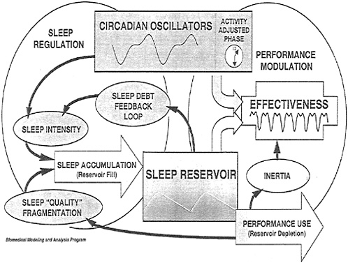
FIGURE 5–1 A schematic of the Sleep, Activity, Fatigue, and Task Effectiveness (SAFTE) model.
SOURCE: Eddy and Hursh (2001). Figure reprinted with permission of Biodynamics and Protection Division, Human Effectiveness Directorate.
voir. The sleep accumulation process is affected by sleep intensity (which is modulated by existing sleep debt and circadian factors) and quality of sleep (which is affected by sleep continuity). Cognitive readiness or effectiveness is predicted based on the level of the sleep reservoir and the time of day (circadian phase), as well as on the potential influence of short-term, postsleep grogginess (referred to as “sleep inertia”).
This model has been implemented through the Fatigue Avoidance Scheduling Tool. This tool is useful for identifying times at which performance might be compromised within a given work/sleep schedule, and it is useful for optimizing schedule development because it allows an operator/planner to ask a series of “what if” questions. For example, as shown in Figure 5–2, a planner can view the predicted effects of 2 days without sleep, and then ask “What if we placed a 4-hour nap during this 40-hour period of otherwise continuous wakefulness?” As the figure indicates, such a napping strategy could offer a 20 percent
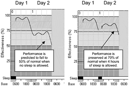
FIGURE 5–2 An example of output from a Fatigue Avoidance Scheduling Tool (FAST) to help predict cognitive effectiveness (%). Example: 2 days with no sleep compared with 2 days with 4 hours of sleep.
improvement in cognitive effectiveness over what would be expected with no sleep at all.
The predictive capability of the SAFTE model has been established by comparing the model’s output with laboratory data collected during various sleep-deprivation studies. For example, model predictions accounted for 89 percent of the variance (degradations) in throughput on serial addition-subtraction across 72 hours of sleep loss in one study, and 98 percent of the variance in throughput on a variety of cognitive tests across 54 hours of sleep deprivation in another (Hursh et al., 2004). Throughput is a combined speed/accuracy measure that on many basic cognitive tests is presumed to reflect the individual’s capacity to perform mental discriminations, react to incoming stimuli, think logically, process information, and comprehend language. Although the tasks on which SAFTE was validated are not typical military tasks, it is assumed that anything that degrades such basic mental facilities would also degrade operationally relevant performance. Future studies will validate the relationship between SAFTE model predictions and decrements in a variety of “real-world” performances.
SAFE
The SAFE model (Belyavin and Spencer, 2004) is similar to SAFTE in that it takes keyboard input about specific work/rest schedules and estimates per-
formance risk based on what is known about the impact of the body’s clock and time since sleep on alertness. In addition, the model accounts for the effects of sleep inertia. SAFE predictions were initially validated via comparisons with laboratory data collected during several studies of sleep deprivation and shift work. Select variables were used from tests of visual vigilance, continuous memory recall, psychomotor tracking, and the Multi-Attribute Task Battery. The results indicated that the basic model effectively predicted group performance on most aspects of these basic tasks.
Later, SAFE predictions were compared with the subjective alertness ratings of a sample of commercial airline pilots across 72 flights (comprised of long-haul international trips). On some schedules, the model predicted mean alertness levels moderately well, with the exception of a sudden increased arousal that occurred at the end of some of the return flights. On other schedules, although the model tracked fatigue-related changes fairly well from the beginning to the end of each flight segment, the model underestimated alertness on outbound flights and overestimated alertness on return flights. Because of these discrepancies, the designers of SAFE performed additional studies and ultimately included prediction modifiers that considered not only the three basic alertness processes (time since sleep, circadian rhythms, and sleep inertia), but added the effects of: (a) multiple flight legs, (b) duration of time on duty, (c) the effects of consecutive and/or long tours of duty, (d) the impact of early report times, (e) the impact of daytime vs. nighttime sleep, and (f) the effects of sleep degradations during on-board rest periods. The addition of these factors substantially increased the predictive accuracy of the model.
Sleep Monitoring
There are two basic approaches for monitoring human sleep. The first, and most accurate, consists of electrophysiological recordings (polysomnograms). The second, and most practical for nonlaboratory settings, consists of activity-based recording (actigraphy).
Polysomnography
Polysomnographic recordings involving the collection of EEG, electromy-ographic (EMG), and electrooculographic (EOG) data from skin-mounted electrodes represent the most accurate way to monitor sleep parameters. For clinical and research purposes, sleep recordings are usually made in a sleep laboratory because of the available level of environmental control and the instrumentation required. EEG data are acquired with silver-silver chloride or gold electrodes attached to the scalp with collodion before the person retires for the night. A minimum of one or two EEG electrodes are attached, along with a mastoid (or ear-lobe) reference, two EOG electrodes, and two EMG electrodes placed underneath the subject’s chin. Amplification of the signals is accomplished via
highly sensitive polygraph amplifiers. The signals are recorded on paper, usually at a speed of 10 mm/sec, or by a computerized system configured for sleep polygraphy. Using standard guidelines, the stages of sleep (stages 1 through 4, REM, awake, and movement) are determined by scoring each epoch (20–30 second period) throughout the record. Most data are scored visually following standardized criteria set forth by Rechtschaffen and Kales (1968). New computer systems have been developed to score the data using mathematical functions but, at present, there is no widely accepted automatic scoring procedure that can take the place of a human sleep scorer.
Polysomnographic sleep evaluation is the “gold standard” in sleep science. These standard recordings have been used for years to document and understand “normal sleep,” as well as to investigate a wide array of clinical complaints, including excessive daytime sleepiness, insomnia, and a number of sleep disorders (Carskadon and Dement, 1994; Dement, 1976; Hauri and Orr, 1982; Roffwarg, 1979). The polysomnographic approach avoids the pitfall of making potentially erroneous assumptions that behaviorally quiescent people are asleep when, in fact, they may simply be lying still. This approach also offers a wealth of information about the depth (or quality) of sleep than cannot easily be determined by other means. However, these advantages are offset by recording and instrumentation requirements that make in-the-field sleep recordings at best, impractical, and at worst, impossible. Although various types of ambulatory physiological recorders are available for nonstandard recording applications, it is unlikely that these will be used on a widespread basis (for large groups of personnel) any time in the near future. First of all, there is significant overhead involved in the attachment and maintenance of suites of recording electrodes and, second, the necessity of downloading and processing the data recorded from large numbers of people on a daily basis is impractical. These are the reasons that actigraphic approaches to estimations of sleep quantity have received increased attention.
Actigraphy
Actigraphs are battery-powered, wristwatch-like units that are programmed via a microcomputer interface. The wristwatch contains a three-axis accelerometer sensor with a preamplifier and filter that are interfaced to a microprocessor by a multiplexed analog-digital converter. The microprocessor uses storage memory to record the collected data. The actigraph is programmed by a microcomputer to start and end data collection at specific time points; the same microcomputer interface is used for periodic data retrieval. Once downloaded, the activity data are processed by analysis software that permits sleep-state scoring, basic statistics, and several forms of visual presentation. Raw graphic depictions of data show different levels of activity using a series of discrete vertical lines of different amplitudes. Typically these graphs are scored by both computer algorithms and a human observer (who checks the computer scoring).
Recent investigations have shown that actigraphy is a fruitful approach to the study of sleep in nonlaboratory settings. Pollak and colleagues (2001) compared actigraphically measured sleep/wakefulness with standard polysomnographic measures and found that actigraphs could correctly identify periods of sleep versus wakefulness. Furthermore, the actigraphs offered a general indication of sleep quality in that lower actigraphic counts corresponded to deeper sleep stages. Monk and colleagues (1999) found that actigraphic recordings were useful for determining the sleep onset and offset of four astronauts engaged in 17-day Space Shuttle missions. There was a high correlation between high activity counts and polygraphically identified light stages of sleep. Wrist actigraphy is a promising monitoring strategy for military field applications—it may be used in numerous operational contexts since the required equipment is small, and it is a measure that can be obtained without disturbing the ongoing mission.
Actigraphy in Combination with Performance Prediction Models
In addition to using actigraphy to record sleep/wake cycles and to estimate sleep quality and quantity, efforts are underway to integrate these data with computerized performance prediction algorithms. One of the best developed of these applications is the Walter Reed Army Institute of Research Sleep Watch Actigraph (Balkin et al., 2000). The Sleep Watch is a wrist-worn device that predicts the wearer’s cognitive performance status based on the level of sleep debt and the phase of the circadian cycle (as calculated via actigraph activity counts). The actigraphically determined sleep/circadian information is fed into an on-board computer model that includes a “charging function” for recuperative sleep, a “discharging function” for the amount of time awake, and a “circadian function” for the specific phase of the wearer’s estimated circadian rhythm. (This is the Sleep Performance Model that was mentioned earlier.) The face of the Sleep Watch contains a “fuel gauge” that displays a performance prediction ranging from 0 to 100 percent of well-rested levels and a simple red-yellow-green analog scale that can alert the wearer of the need to acquire additional sleep. The Sleep Watch is still under development to include individual differences in sleep needs and performance capabilities and the effects of time-zone transitions and variations in light exposure levels (which influence circadian adaptation). The Sleep Watch predictions have been validated to some extent on numerous tasks collected during sleep-deprivation and sleep-restriction studies in the laboratory, and further work along these lines is currently underway. There are plans to employ the Sleep Watch in upcoming field exercises so that Watch-based alertness measures can be correlated with aspects of real-world performance. Once this tool is optimized, it will offer a fleld-useable method for monitoring operator fatigue levels and for warning wearers of impending cognitive performance failures associated with insufficient sleep.
Spontaneous Electroencephalography
Operational personnel are required to perform even if they are fatigued. In these situations it is desirable to have the capability to continuously monitor the cognitive status of combat service members without interfering with the accomplishment of their primary mission. Although there are cognitive tests that are exquisitely sensitive to the presence of fatigue stemming from sleep loss, circadian disruptions, and a host of other factors, it is difficult to continuously administer such tests on an hourly or daily basis without disrupting job performance. Thus a more unobtrusive monitoring strategy is required. One possibility is to take advantage of the spontaneous EEG because the data can be obtained and to some extent, examined, in real time, and the results are considered to be objective, valid, and related to operational readiness (Caldwell et al., 1994, 1997, 2002). As mentioned earlier, one of the key advantages of the EEG is that it is a psychophysiological measure that can be collected without interfering with the primary task.
EEG Measures
EEG signals are detectable from a variety of scalp locations using small electrodes (8 mm) that are attached with tape, collodion, or paste and are filled with an electrolyte solution. Electrical activity is then recorded with two or more electrodes (depending on how much of the brain’s surface activity is of interest) by amplifiers capable of amplifying 5- to 100-μV signals that oscillate at 0.5 to 100 Hz. Amplified data are usually stored on an ambulatory or laboratory recording device and/or displayed on an ink-writing polygraph or a computer cathode-ray tube. Recorded data may be scored via visual inspection or by computerized procedures, such as power spectral analysis. In either case, the recording is usually reduced to a table of values that represents the relative amount of EEG falling within each of four distinct bands: delta (1–3.9 Hz), theta (4–7.9 Hz), alpha (8–13 Hz), and beta (>13–20 or 30 Hz). Also, the dominant or peak frequency observed during specified time frames can be determined. The normal adult waking EEG contains each of these different rhythms, but the most easily recognizable one is the alpha rhythm that is blocked by opening the eyes, cognitive activation, sudden arousal, and sleep. Beta waves also contribute substantially to the normal awake EEG, but slower activity (below 8 Hz) is usually predominant only during sleep (Cooper et al., 1980) or under conditions of severe drowsiness.
Numerous studies have established the sensitivity of EEG activity to work-related stressors (e.g., sleep deprivation). For instance, Caldwell and colleagues (1996), Comperatore and colleagues (1992), Lorenzo and colleagues (1995), and Pigeau and colleagues (1987) have all shown that slow-wave EEG activity (i.e., delta and theta) is significantly elevated by even moderate sleep loss (i.e., sleep loss resulting from 18–20 hours of continuous wakefulness), and research has repeatedly established that the fatigue from insufficient sleep affects both EEG
activity and nonconcurrently measured flight performance (Caldwell et al., 2000a, 2000b, 2003; LeDuc et al., 2000). Based on these findings, it is reasonable to suspect that EEG data should show a predictive relationship to operationally relevant cognitive performance. However, while several investigators have offered evidence that EEG is useful for describing an operator’s general status during tasks such as flights (Blanc et al., 1966; Caldwell et al., 2002; Howitt et al., 1978; Maulsby, 1966) and driving simulations (Balkin et al., 2000), correlations with actual task performance are often weaker than desired except in cases of extreme drowsiness.
It should also be noted that while a strong link between EEG and drowsiness has been demonstrated, there have been a host of problems related to real-time EEG monitoring in operational environments. These include the necessity of highly trained technicians, cumbersome and fragile instrumentation, and a rather time-consuming preparation period for EEG recordings. Additionally, the acquired data is susceptible to contamination by physiological and instrumental artifacts, such as movements, muscle activity, eye blinks, or heartbeats. Also, while much progress has been made in overcoming these obstacles, there is no general consensus on what aspects of EEG are sufficiently sensitive and reliable to serve as general indicators of fatigue (Gevins et al., 1995).
Nonetheless, the utility of EEG continues to be explored because of a logical association between brain electrical activation and brain function. New analysis routines, better equipment, automated artifact-correction algorithms, and new types of high-impedance electrodes will ultimately make widespread real-time monitoring of cognitive readiness (via measures of brain activity) feasible in field settings. As Gevins and colleagues (1995) have pointed out, “the EEG has one or more advantages over other [central-nervous system monitoring] approaches, including millisecond temporal resolution, complete harmlessness and noninvasiveness…and [the fact that] EEG is the only brain imaging modality suitable for use in operational environments or from ambulatory subjects.”
A SMART HELMET, designed to provide inputs for 32 channels of low-level electrophysiological signals, including EEG, EOG, EMG, and heart activity (electrocardiogram [ECG]) (Gevins et al., 1995) was once available. Additional channels offered the capability to record measures such as respiration, blood volume, skin conductance, and head and body movements. In conjunction with preamplifier circuits and electrically driven shielding built directly into a flight helmet, a stretchable fabric hat was used for rapidly positioning EEG, EOG, and EMG electrodes on the head and face for recording data. The SMART HELMET successfully collected data from subjects in a centrifuge, airplane, and car with the use of ruggedized laptop personal computers (Gevins et al., 1995). However, the requirement for low-impedance sensors (e.g., skin preparation, electrolyte solution) limited the usefulness of such a technique, and at present, the SMART HELMET is no longer available (although it may pave the way for similar, more modern technology). In all probability, newer devices
will strive to actually embed EEG (and possibly EOG) sensors into an integrated helmet configuration (as opposed to using a stretchable hat worn under the helmet liner). In this way, physiological data reflecting pilot alertness may soon be available every time a pilot dons his or her flight gear. A small Ohio-based company (SRICO) is currently working to overcome one of the major obstacles to using such an embedded-sensor approach by developing a high-impedance, dry-contact EEG sensor (the Photrode) (Kingsley et al., 2003) that can be mounted inside of a flight helmet or a duty uniform. Photrode is still under refinement, but it should be widely available within 2 to 3 years. Once this technology can be coupled with high-capacity recording devices equipped with software routines that can remove contaminating artifacts from movements, muscle tension, eye blinks, and heart beats, the feasibility of continuous real-time EEG monitoring in the field will improve. Ultimately these data could be used to feed performance prediction models, such as the one currently used for Sleep Watch. This should be an available option in 5 to 7 years.
EEG Measures Combined with Performance Measures
Sleep-watch measures, EEG assessments, or other physiological parameters may be the ultimate strategy to monitor the cognitive states of ground combat service members, but combined physiological and performance measures may be a better alternative for vehicle operators. Caldwell and Roberts (2000) have shown that objective, integrated flight-performance assessments are sensitive to the impact of fatigue and specific types of antifatigue medications, and others have noted the sensitivity of EEG measures to the effects of fatigue in a variety of situations (Balkin et al., 2000; Hossain et al., 2003; Miller, 1997). However, little work has been done to develop status predictors that rely on the integrated combination of continuous performance assessments and concurrent evaluations of physiological status. Petit and colleagues (1990) have shown that steering wheel functions and EEG alpha power correlate, and de Waard and Brookhuis (1991) found that the standard deviation of steering wheel movements increased and the number of steering wheel reversals per minute decreased in conjunction with a gradual decrease in relative EEG energy. Brookhuis and de Waard (1993) identified the co-occurrence of changes in physiology and in behavior and demonstrated the feasibility of monitoring a vehicle operator’s status by monitoring driving performance. In part, this led Brookhuis (1995) to contend, “Measuring physiological parameters could be considered from the point of view of validating non-obtrusive in-vehicle measures that might be used to monitor driver state continuously through vehicle parameters.” However, it may be unwise to rely on either type of measure (physiological vs. performance) alone in situations where both could be integrated to predict impending cognitive decrements. Integrated monitoring of both categories of measures appears to be a more fruitful approach for future monitoring efforts.
Electrooculographic and Other Eye-Movement Measures
The sensitivity of the oculomotor control system to fatigue, boredom, and lapses in attention has also been noted. The process of monitoring eye movements, EOG, is concerned with measuring fluctuations in electrical potentials during movement of the eyes. EOG measurements have been used in a wide range of applications, such as the recording of REM during sleep research (Andreassi, 1989). It has also been found that long-duration eye closures during blinking are related to reduced alertness (Stern, 1980).
General Ocular Measures
Stern and Ranney (1999) identified a series of oculomotor measures that may be potentially useful for detecting lapses in attention. The first of these is saccadic eye movements. These are the types of eye movements that quickly transition the eyes from one point of focus to another, such as when reading text. Surrounding the occurrence of the saccade is a brief period during which information intake is inhibited. The latency between stimulus presentation and the saccade, the saccade duration, and the distance of the saccade have been suggested as indicators of fatigue. Although saccades are initiated under voluntary control, once they are underway their speed is not within the individual’s control. Fatigued conditions can cause a longer latency period, reduce the velocity of the saccade, or result in saccades that either undershoot or overshoot the target. Russo and colleagues (2003) found that saccadic velocity is particularly sensitive to an increase in sleepiness in response to prolonged periods of partial sleep deprivation.
Another type of oculomotor measure is blinks. Eye blinks can be measured in terms of blink frequency, timing in respect to stimulus presentation, and duration of the closing and reopening movement of the eyelid. It has been found that fatigue can cause smaller-amplitude eye blinks. Similarly, the frequency of eyelid closures has been shown to increase under fatiguing conditions.
A third type of oculomotor measure is pupil diameter. Stern and Ranney (1999) point out that a decrease in pupil diameter or a slow fluctuation in pupil diameter coincides with feelings of fatigue. Similarly, Russo and colleagues (1999) found that decreases in saccadic velocity and increases in pupil constriction latency correlated with an increase in the rate of crashes seen in simulated driving conditions during periods of sleep deprivation.
PERCLOS
Research in the area of slow eye closures has given rise to PERCLOS, a measure defined as the “percentage of time that the eyes are 80% to 100% closed over a defined time interval” (Wierwille, 1999). This type of measurement has the advantage of being physiologically based, and it has a great deal of face validity since drooping or slow eyelid closures are not usually seen in alert
individuals. Furthermore, performance decrements on a variety of tasks are virtually certain to occur when slow eye closures impair an individual’s ability to gather visual information. Early research developed algorithms that combined estimations of ocular measures (PERCLOS) with a direct measurement of performance (Wierwille, 1999). Dinges and colleagues (1998) have determined that there is a high degree of coherence between PERCLOS and performance lapses on the psychomotor vigilance test (an accepted test of fatigue). At present, PERCLOS is a labor-intensive monitoring technology that involves the human scoring of eye closures from video footage of volunteers’ faces. However, a direct on-line PERCLOS system that uses infrared illumination to compare retinal reflections against a dark background and automatically calculates a real-time PERCLOS value is being developed. This has led to a dash-mounted Copilot, a “low-cost drowsiness monitor intended for use in commercial operations involving nighttime driving” and “designed for robust operation in a heavy truck environment” (Grace and Steward, 2001). This is a technique that may one day be useful for monitoring vehicle operators in field environments, but the equipment involved makes it unlikely that PERCLOS can be employed to assess the operational readiness of ground combat service members.
Other Ocular Measures
There are a number of commercial products available that measure fatigue. One such device is the FIT Fatigue Analyzer (PMI, Inc., Rockville, Maryland). This particular device can assess the degree of fatigue by analyzing involuntary pupillary responses to brief flashes of light and by analyzing eye movements in response to moving light targets. Measures of pupil size, constriction latency, constriction amplitude, and saccadic velocity are combined into a weighted score that purportedly assesses fatigue levels. There are efforts underway to miniaturize this device for operational use.
Similarly, MTI Research Inc. has developed a device designed to detect and track fatigue using eye-blink analysis. Using optical electronics, the Alertness Monitor determines the level of alertness or drowsiness by measuring the ratio of eyelid closures to eyelid openness. Mounted unobtrusively on safety glasses, the research models emit an infrared beam along the axis of the eye blink where the beam cannot be broken by the eyelashes during an eye blink and will not shine directly into the operator’s eye (Dinges et al., 1998). Meanwhile, IM Systems, Inc. has developed the Blinkometer, an ambulatory device that records eye blinks using an algorithm reported to be sensitive to drowsiness. The Blinkometer can record in one of two modes: either blinks per minute or the intervals between blinks. The device detects blinks using a sensor attached to the outer canthus of one eye with a double-sided adhesive disk and a small recording device clipped to the operator’s person. IM Systems reports that by using a fairly straightforward algorithm (the fewer the number of blinks, the greater the level of drowsiness), the Blinkometer detects a decreased alertness within 20 to 30
seconds (Dinges et al., 1998). Both the Alertness Monitor and the Blinkometer were tested for validation, alongside PERCLOS, by comparison with lapses in a psychomotor vigilance task. While the “result for PERCLOS was uniformly high coherence” to these attention lapses as seen in the psychomotor vigilance task, the result for the Alertness Monitor was “at the other end of the spectrum, averaging the lowest bout-to-bout coherence for lapse frequency” (Dinges et al., 1998). The Blinkometer had a moderate bout-to-bout coherence for lapse frequency, but was problematic due to difficulties with the unit’s data storage and retrievability functions (Dinges et al., 1998). While most of these technologies have potential, Dinges and colleagues (1998) pointed out that “more validation studies of this type are needed to sort out from the wide variety of biobehavioral fatigue monitors those that have the highest validity and reliability for predicting actual hypo-vigilance performance.”
Another device, originally developed as a home sleep-monitoring system, also shows promise in this area. The Nightcap, developed by Healthdyne Technologies, detects eyelid movements through a small, piezoelectric film sensor, which is attached to the upper eyelid. This method allows the detection of both active movements of the eyelid and passive movements caused by movement of the eyeball. The Nightcap detects decreases in vigilance as lowered levels of eyelid movements (Stickgold et al., 1995). Using the Nightcap, Stickgold found decreased eyelid movements during periods of decreased vigilance resulting from inadequate sleep on the previous night. Stickgold and colleagues (1995) concluded that “the Nightcap would appear to be potentially useful for the real-time monitoring of vigilance in a variety of work environments.”
A similar method for measuring eyelid movements originally developed by Evinger and colleagues (1991), in which a very small piece of insulated wire coil is taped to the upper eyelid while the operator sits in a 3-D magnetic field, has been used by Leder and colleagues (1996) to study the relationship between eyelid activity and alertness/vigilance. They report that the magnetic sensor followed the wire coil well and responded to every blink. Additionally, this method was able to distinguish spontaneous blinks from vertical lid saccades and horizontal eye saccades. As a result, Leder and colleagues (1996) “…expect to be able to unobtrusively collect spontaneous eyelid activity in ambulatory subjects engaged in their routine activities,” including “changes in blink rate associated with fatigue and loss of alertness or vigilance.”
OTHER CENTRAL NERVOUS SYSTEM MONITORING TECHNOLOGIES
In addition to the technologies described above, there are other methods of studying central nervous system changes that may offer information about cognitive status. However, none of these are suitable for field applications in which continuous, real-time assessment is the goal.
Positron Emission Tomography
Wu and colleagues (1991), with the use of positron emission tomography (PET), found that about 32 hours of total sleep deprivation decreased metabolism in the thalamus, basal ganglia, white matter, and cerebellum. Sleep deprivation further reorganized regional cerebral metabolic activity by decreasing temporal lobe activation and increasing activity in the visual cortex. As a result, visual vigilance on a continuous performance test was degraded. Thomas and colleagues (2000) reported that 24 hours of sleep deprivation produced significant decreases in relative regional glucose metabolism in the thalamus and prefrontal and posterior-parietal cortices of the brains of 17 volunteers. Once again, both alertness and cognitive performance declined in conjunction with changes in brain activity. Although not within the context of sleep deprivation, Pietrini and colleagues (2000) have asserted that PET is also useful for tracking the changes in neural activity that accompany the cognitive declines associated with Alzheimer’s disease. Scans of patients revealed a progressive decline in the magnitude of brain response to audiovisual stimulation with progressive worsening of cognitive dementia. Thus PET offers important information about the levels of brain activation that underlie cognitive performance; however, the instrumentation and testing requirements for this method cannot be met in an operational context.
Functional Magnetic Resonance Imaging
Downing and coworkers (2001) have used neuroimaging techniques, such as functional magnetic resonance imaging (fMRI) and magneto-EEG, to identify the neural substrates of visual attention. The results indicated that these techniques are able to identify markers of face processing and place processing. Likewise, Rees (2001) has addressed the relationship among selective attention, neural activity, and visual awareness through fMRI research. Portas and coworkers (1998) have used fMRI to help delineate the role of specific brain areas in attention and arousal (both of which are important for cognitive performance). The authors identified differences associated with the performance of an attention task as a function of arousal decrements. Sleep deprivation produced an increase in attention-related thalamic activity compared with what was observed after administration of caffeine to improve arousal. Drummond and colleagues (2000) observed that while sleep deprivation increased subjective sleepiness, prefrontal cortex activation was actually more responsive after 1 night of sleep deprivation than after normal sleep. Sleep deprivation also impaired performance on a free-recall task, but it was observed that free-recall was improved in subjects with greater parietal lobe activation. The prefrontal cortex results from this study appear to partially agree with a later finding that sleep deprivation was associated with greater activation in the bilateral prefrontal cortex and parietal lobes during the performance of verbal learning and divided attention tasks (Drummond and Brown, 2001). However, the fact that sleep deprivation led to
decreased activity in these same areas during the performance of an arithmetic task suggested that the brain’s response patterns are dependent on the type of cognitive processing required. Clearly, fMRI can offer useful information about the brain areas involved in specific types of task processing, as well as about the effects of fatigue on basic central nervous system functioning. However, as is the case with PET, fMRI is not suitable for field applications.
Transcranial Doppler Sonography
Transcranial doppler (TCD) sonography is a method for noninvasively monitoring cerebral blood flow levels that can serve as an indicator of metabolic activation in the brain. TCD has been used in the study of vigilance, as well as to understand the cerebral processes underlying various cognitive tasks (Hollander et al., 2002). For instance, Hitchcock and colleagues (2003) found that there were performance-related changes in right hemisphere blood flow as a result of manipulations that increased or decreased the demands of a 40-minute, simulated air traffic control task. Other investigations have demonstrated the potential utility of TCD in understanding the cerebral organization of cognition. However, Stroobant and Vingerhoets (2000) indicated that additional standardization of procedural methodologies will be required before the full utility of TCD can be realized. In any event, it remains unlikely that such a technique will ever be feasible for monitoring foot-soldier status in the field.
HEART-RATE MEASURES
Although heart-rate measures typically have not been used to assess aspects of cognitive readiness associated with fatigue or sleepiness, heart rate has often been employed to assess other aspects of operator state. The heart is innervated by both the sympathetic and the parasympathetic nervous systems and, as such, it is influenced by higher cortical centers. The sympathetic nervous system increases the firing rate of the heart’s pacemaker and also modulates the constriction and dilation of the blood vessels. The parasympathetic nervous system inhibits the firing rate of the pacemaker cells via the vagal nerve, and this reduces heart rate. Of course heart rate has long been used as an indicator of physical effort, but it has also been proven useful for studying mental effort and other aspects of psychological and cognitive status. Thus the monitoring of heart rate as an indicator of cognitive stress may be useful for optimizing task demands with the aim of avoiding levels of cognitive fatigue that could lead to a breakdown in alertness or performance capacity.
As described by Caldwell and coworkers (1994), numerous studies have found systematic relations between cognitive demands and heart rate in both laboratory and real-world environments (for reviews, see Kramer, 1991; Roscoe, 1992; and Wilson and Eggemeier, 1991). In addition, the operational relevance of heart-rate measures has been well established in demanding performance con-
texts, such as flying combat missions (Lewis et al., 1967), flying surface-attack training missions (Comens et al., 1987; Wilson, 1993), flying aircraft test missions (Roscoe, 1980), and landing at different airports (Nicholson et al., 1970; Ruffell-Smith, 1967). Heart-rate changes have been shown to discriminate between pilot vs. copilot flying (Hart and Hauser, 1987; Kakimoto et al., 1988; Roscoe, 1978) and flying in the lead versus the wing position (Wilson et al., 1987). Simulated flight studies have also reported increases in heart rate associated with increases in task difficulty (Lindholm et al., 1984; Opmeer and Krol, 1973; Wierwille and Connor, 1983).
Heart-rate variability (HRV) is also a sensitive indicator of task demands. Several studies have reported decreases in HRV with increasing cognitive workload. Specifically, it has been found that after performing a spectral analysis on the ECG signal, an examination of the midfrequency band (the 0.10 Hz component) offers information about the amount of mental effort that has to be invested to meet the task demands (Mulder, 1992; Mulder and Mulder, 1980). The tasks evaluated have included simulated and actual flight (Lindholm et al., 1984; Lindqvist et al., 1983; Opmeer and Krol, 1973; Sekiguchi et al., 1979; Wilson, 1993). Opmeer and Krol (1973) reported that HRV and respiration were sensitive to simulated flight task demands. Itoh and coworkers (1989) found HRV in the 0.10 band to decrease during take off and landing (high workload) when compared with cruise segments (low workload).
Recently, researchers have also found that HRV in the high-frequency band (0.15–0.40 Hz) can be used to predict cognitive performance. Tasks that have been examined include the Stoop color-word conflict task, continuous performance tasks, and working memory tasks. Johnsen and coworkers (2003) found that dental phobics with higher HRV had faster reaction times to incongruent color and threat-related words than did dental phobics with lower HRV. Importantly, Hansen and colleagues (2003) recently reported the results of a study in a military sample. Naval cadets in the Royal Norwegian Navy were divided in to high- and low-HRV groups based on their resting HRV. These researchers reported that those individuals with high HRV performed better on tasks that used executive functions, such as working memory, compared with those with low HRV.
Since monitoring ECG is in many ways easier than monitoring EEG (due to greater signal strength and subsequent improvement in signal-to-noise ratio), the collection and analysis of heart rate and HRV is actually quite feasible at this point. With the introduction of high-impedance sensors that can be mounted in standard clothing, it is likely that these cardiac variables will soon be routinely monitored, at least in some specialized training environments. After all, commercially available systems consisting of chest straps and wrist-worn receivers (such as the Polar systems) are already widely used by athletes.
OTHER MEASURES
It should be noted that there are several other types of psychophysiological measures besides EEG, EOG, and ECG that hold promise for assessing aspects of operator cognitive status. For instance, changes in body temperature not only reflect different levels of physical energy expenditure and changing environmental conditions, but also fluctuations in psychological arousal. Differences in muscle activation measured by EMG recordings can indicate increased or decreased physical activity or elevations or reductions in psychological tension. Fluctuations in respiration rate can suggest either changes in physical energy expenditure or changes in mental stress. These and other psychophysiological measures are not widely used for the assessment of cognitive readiness per se, but they can provide insight into psychological or work-related factors that ultimately can influence operator status. Since all of them can be assessed via non-intrusive, skin-mounted sensors, their potential utility for future applications should not be dismissed; however, each measure poses different challenges in terms of both recording and analysis. A complete discussion of all of the available psychophysiological measures is beyond the scope of this report, but interested readers may wish to consult one of more of the authoritative texts that have already compiled and synthesized detailed information on this topic, such as Andreassi (1989), Cacioppo and Tassinary (1990), and Coles and colleagues (1986).
Head-Position Monitoring
Head-position monitoring, although not considered psychophysiological, is another method that has been thought to hold promise as a measure of fatigue. One of the self-assessment indicators of fatigue that many operators watch for is increased head bobs or other involuntary movements that occur due to loss of neck muscle tone. Stern and Ranney (1999) suggest that an increase in the amount of reactive head movements (as opposed to eye movements) when attempting to react to some event may be a sign of fatigue. This is thought to indicate an increase in passive responding. Some research suggests that there may in fact be a relationship between micromotion of the head and fatigue and, because of this, Advanced Safety Concepts, Inc. has developed the Proximity Array Sensing System (PASS). This apparatus records the x, y, and z coordinates of the head, using an array of three capacitive sensors that are mounted overhead in a vehicle or other fixed location. The position of the head is triangulated by determining the proximity of the head to each capacitive sensor through partial blocking of the sensing fields. It is hypothesized that changes in head-movement patterns may indicate fatigue onset. It has been reported that in laboratory tests, “the PASS system has detected changes in head position as little as 0.01,” while providing absolute x-y-z resolution of head position to about 0.1” (Dinges et al., 1998). “Beta” systems that can be used in simulator research and in real-world trials are under development (Kithil et al., 2001).
Voice Analysis as a Monitoring Approach
For some time the law enforcement community has utilized methods of speech examination as a source of information on intoxication from alcohol and drugs. In the highly publicized Exxon Valdez accident, speech analysis was used to help determine that the responsible individual’s judgment was impaired by alcohol (Brenner and Cash, 1991). Brenner and Cash (1991) note that alcohol ingestion produces slowed speech, speech errors, misarticulation of difficult sounds, and changes in vocal quality. It is possible that speech analysis might also be useful during fatigued conditions, as presumably many aspects of fatigued speech would be similar to speech under the influence of alcohol (a depressant of the central nervous system).
Brenner and colleagues (1994) have suggested that analyzing an operator’s physiological state with speech-based analyses would be useful in the aerospace environment since speech analysis, unlike many other measures, is unobtrusive and does not require the pilot to be attached to any equipment. Speech measurements can be obtained through preexisting, on-board communications equipment (Brenner et al., 1994). These authors have identified six aspects of speech that may be applicable for determining psychological state (at least stress and possibly fatigue): (a) speaking fundamental frequency (pitch), which increases under stress; (b) speaking rate, which increases under stress; (c) vocal intensity (loudness), which increases in decibels as a function of the increased thoracic air pressure that occurs under stress; (d) vocal jitter, a subtle measure of the minute changes in the period of successive fundamental frequency cycles, which decreases in response to stress; (e) vocal shimmer, which is analogous to jitter and reflects the cycle-by-cycle differences in vocal intensity; and (f) derived speech measure, which combines properties of several speech measures and may provide a more sensitive indicator of stress. It was found by Brenner and coworkers (1994) that speaking fundamental frequency, vocal intensity, and speaking rate all increased in response to changing workload demands.
Johannes and coworkers (2000) point out that vocal pitch is affected by changes in autonomic nervous system arousal and that, in general, an increase in fundamental pitch is associated with emotional excitation. However, these authors go on to point out that useful voice-based state predictions are hampered by considerable individual differences in fundamental pitch, as well as by differing reactions to stressful events. In addition, some type of initial state calibration is required to differentiate the effects of stress versus fatigue. Whitmore and Fisher (1996) found that speech signals (word duration and fundamental frequency) recorded from aircrews in B-1B long-range bomber simulators tended to fluctuate parallel to the circadian cycle, like subjective and cognitive performance, during sleep deprivation. Word duration lengthened and fundamental frequency decreased as a function of fatigue. Griffin and Williams (1987) determined that an increase in peak amplitude and a decrease in word duration during conditions of increased workload were indicative of task complexity.
Additional research on the manner in which speech degrades under fatigued conditions may enable the development of new strategies to monitor both cognitive load and fatigue. However, it remains to be seen whether this technology will ever be applicable to military or other operational settings. In fact, a 1990 review of 50 years of research on voice analysis techniques indicated that while this is a promising area of research, the results have not shown voice analyses to be reliable techniques for determining the type or degree of reaction to stress or workload in operational settings (Ruiz et al., 1990). Studies on the utility of voice analysis for the prediction of changes in cognitive readiness are virtually nonexistent.
Self-Assessment
Standardized assessments of basic cognitive skills and periodic self-ratings of alertness and performance capabilities can provide valuable insight into the functional status of personnel as long as basic standardization (and a few other criteria) are met. With regard to cognitive assessments, Santucci and colleagues (1989) point out that cognitive tests are useful for monitoring the impact of environmental stressors and for evaluating the information-processing capabilities of individuals. However, these authors also caution that it is difficult to interpret the results of mental performance tests in uncontrolled, rapidly changing environments, across individuals who may show wide (and unknown) individual differences, and in circumstances in which the effects of practice cannot be controlled. Also, deciding which tests are best for each military job specialty may be difficult. Psychomotor tasks may seem optimal for monitoring the performance status of vehicle operators, whereas visual-attention tasks may appear to be a better choice for predicting decrements in radar, sonar, and radio operators. However, validation procedures must substantiate such assumptions, and there are no doubt extensive interactions among test requirements and test characteristics that will complicate the selection of the most appropriate choice for each occupational specialty. Subjective self-ratings appear more straightforward as long as their implementation is feasible and there is little chance that the personnel being assessed will be motivated to over-report or under-report the subjective symptoms of interest. For instance, Dorrian and coworkers (2000) have reported that research subjects were globally able to self-assess neurobehavioral performance decrements attributable to increases in fatigue with a high degree of accuracy. Gillberg and colleagues (1994) likewise found that three different subjective sleepiness scales were highly correlated with performance on a visual vigilance and a reaction-time task over the course of a night shift. More relevant to the military operational context, Caldwell and coworkers (2003) reported that preflight self-ratings of fatigue (from the Profile of Mood States) correlated highly (−0.72) with pilots’ abilities to accurately complete simulator flight maneuvers during a 37-hour period of continuous wakefulness. In addition, as described in Chapter 3, self-assessments have proven useful for predicting the lev-
els of optimal physical performance and the extent of performance deteriorations that would be of concern to combat service members engaged in demanding physical tasks. Thus self-ratings of operational status deserve serious consideration for their potential usefulness in status monitoring.
SUMMARY
No doubt there are strategies under development (and under refinement) that may contribute significantly to a further understanding of the basis of cognitive processing, as well as to the effects of fatigue, workload, and other factors that influence human performance. In all probability most will be useful only in laboratory environments or in fixed-based operational facilities (such as posts in which radar and sonar equipment are monitored or stations from which remote-controlled vehicles are piloted) where complex equipment can be housed, lengthy recording procedures can be conducted, and rigid controls can be maintained. Only a small subset of the strategies will likely be suitable for operational settings.
Based on a general review of the literature, it appears that the most promising techniques for accomplishing real-time, continuous assessments of foot-soldier cognitive readiness in military field settings are: (1) actigraphy based, or (2) EEG based, although neither technique is currently ready for widespread application. As noted, the Walter Reed Army Institute of Research has made substantial progress in the development and validation of an actigraph-based, sleep/fatigue monitor that could be worn like a wristwatch in almost any environment. This device may be available by 2005. Concurrent work with high-impedance EEG and ECG electrodes will soon make it possible to continuously record brain activity, heart-rate data, and other electrophysiological parameters and, as noted above, both the EEG and ECG offer useful information about operator status. However, once these new sensors are sufficiently refined, work will remain in terms of mounting them in combat helmets or integrating them into combat clothing. Speech-pattern analysis at one time seemed to hold promise for the future since there is a fair amount of verbal radio communication in the modern operational environment, but the work on this particular measure has not been particularly encouraging.
The most promising techniques for accomplishing real-time, continuous evaluations of the operators of military vehicles; the personnel responsible for manning radar, sonar, or other monitoring equipment; and those whose jobs consist of interfacing with computers and communications devices are: (1) EEG based, or (2) eye-movement based. The recording and evaluation of EEG activity becomes much more straightforward in settings in which operators are physically stationary and quiet because muscle and movement artifacts are attenuated. Furthermore, military aviators are required to wear flight helmets in which newly developed, high-impedance sensors could be mounted. Eye movement parameters (i.e., PERCLOS) have already proven feasible for the detection of
changes in truck driver alertness, and efforts are underway to establish an automated PERCLOS that could be used in aviation settings. Since many eye monitoring systems require the use of cameras that are aimed at the faces of the operators, this is a technology that is clearly more applicable for stationary operators who are already staring straight ahead (at least most of the time) in order to complete some type of monitoring or computer-based task.
Questions about where these new monitoring approaches will be implemented are best considered first by assessing the feasibility of using them in specific environments (as noted above), and second by performing an analysis of the cost of the technology versus the cost of the mishap that the technology would be expected to prevent. Obviously, it is likely to be quite expensive to put some of the newest and most complicated monitoring devices in the hands of every foot soldier or to mount them in every military vehicle, and this in and of itself will pose a substantial barrier to widespread implementation. Thus a jeep driver or a member of a rifle platoon probably will not see the common use of operational alertness monitors for several years after such monitors first become available because of the initial expenses. Furthermore, a performance failure on the part of such individuals is unlikely to be a multimillion dollar catastrophe, so it would ultimately take the military years to reap sufficient savings from the technology to justify implementation in these segments of the overall force structure.
The pilot of a B-2 bomber, however, or those operating other highly complex modern aircraft may be among the first to benefit from newly developed status-monitoring approaches because there are relatively few of these aircraft, and the cost of losing even one would be significant by any standard. Each B-2 aircraft costs more than $1 billion, and the expenses likely to result from a single B-2 air mishap would no doubt be far greater depending on what type of munitions were on board and what the aircraft crashed into during the mishap. On top of these considerations is the fact that B-2s are long-range, two-crew bombers in which aircrew fatigue is known to be an operational hazard (some missions extend well beyond 33 hours of continuous flight time). In light of these facts, automated, onboard alertness monitors would be an obvious choice for fulfilling a much-needed fatigue countermeasure role. Therefore, the costs associated with instrumentation of such a platform are easily justifiable based on the aircraft’s mission and the savings that would result from the prevention of even a single mishap. Such considerations and calculations will no doubt be applied to every potential site for future monitoring applications, at least until a relatively inexpensive and easy solution to the general status monitoring problem is found.
While the search is underway, individuals and their commanders will be forced to rely upon the same types of general alertness predictions (based on group data) and the same subjective impressions about “go” and “no-go” status that have been used for years. A great deal of progress has been made toward helping the armed forces address fatigue-related cognitive decrements once they have been identified. However, highly reliable, efficient, and cost-effective
technological means of initially detecting and predicting those decrements remain to be developed.
REFERENCES
Agnew HW Jr, Webb WB, Williams RL. 1967. Comparison of stage four and 1-REM sleep deprivation. Percept Mot Skills 24:851–858.
Åkerstedt T. 1988. Sleepiness as a consequence of shift work. Sleep 11:17–34.
Åkerstedt T. 1995. Work hours, sleepiness and the underlying mechanisms. J Sleep Res 4:15–22.
Åkerstedt T, Gillberg M. 1982. Displacement of the sleep period and sleep deprivation. Hum Neurobiol 1:163–171.
Åkerstedt T, Kecklund G, Knutsson A. 1991. Spectral analysis of sleep electroencephalography in rotating three-shift work. Scand J Work Environ Health 17:330–336.
Andreassi JL. 1989. Psychophysiology: Human Behavior and Physiological Response. 2nd ed. Hillsdale, NJ: Lawrence Erlbaum Associates.
Angus RG, Heslegrave RJ. 1985. Effects of sleep loss on sustained cognitive performance during a command and control simulation. Behav Res Methods Instrum Comput 17:55–67.
Balkin J, Thorne D, Sing H, Thomas M, Redmond D, Wesensten N, Williams J, Hall S, Belenky G. 2000. Effects of Sleep Schedules on Commercial Motor Vehicle Driver Performance. Federal Motor Carrier Safety Administration. DOT-MC-00–133. Washington, DC: U.S. Department of Transportation.
Belenky G, Penetar DM, Thorne D, Popp K, Leu J, Thomas M, Sing H, Balkin T, Wesensten N, Redmond D. 1994. The effects of sleep deprivation on performance during continuous combat operations. In: Marriott BM, ed. Food Components to Enhance Performance. Washington, DC: National Academy Press. Pp. 127–135.
Belyavin AJ, Spencer MB. 2004. Modelling performance and alertness: The QinetiQ approach. Aviation Space Environ Med 75:A93-A103.
Blanc C, LaFontaine E, Medvedeff M. 1966. Radiotelemetric recordings of the electroencephalograms of civil aviation pilots during flight. Aerospace Med 37:1060–1065.
Bonnet MH. 1994. Sleep deprivation. In: Kryger MH, Roth T, Dement WC, eds. Principles and Practice of Sleep Medicine. 2nd ed. Philadelphia: W.B. Saunders. Pp. 50–67.
Brenner M, Cash JR. 1991. Speech analysis as an index of alcohol intoxication—The Exxon Valdez accident. Aviat Space Environ Med 62:893–898.
Brenner M, Doherty ET, Shipp T. 1994. Speech measures indicating workload demand. Aviat Space Environ Med 65:21–26.
Brookhuis K. 1995. Driver impairment monitoring by physiological measures. In: Hartley L, ed. Fatigue and Driving: Driver Impairment, Driver Fatigue and Driver Simulation. Bristol, PA: Taylor & Francis. Pp. 181–188.
Brookhuis KA, de Waard D. 1993. The use of psychophysiology to assess driver status. Ergonomics 36:1099–1110.
Cacioppo JT, Tassinary LG, eds. 1990. Principles of Psychophysiology: Physical, Social, and Inferential Elements. New York: Cambridge University Press.
Caldwell JA, Ramspott S. 1998. Effects of task duration on sensitivity to sleep deprivation using the multi-attribute task battery. Behav Res Methods Instrum Comput 30:651–660.
Caldwell JA, Roberts KA. 2000. Differential sensitivity of using simulators versus actual aircraft to evaluate the effects of a stimulant medication on aviator performance. Mil Psychol 12:277–291.
Caldwell JA, Wilson GF, Cetingue M, Gaillard AWK, Gunder A, Lagarde D, Makeig S, Myhre G, Wright NA. 1994. Psychophysiological Assessment Methods. AGARD-AR-324. Neuilly-Sur-Seine, France: North Atlantic Treaty Organization.
Caldwell JA, Caldwell JL, Crowley JS. 1996. Sustaining helicopter pilot alertness with Dexedrine during sustained operations. In: Advisory Group for Aerospace Research and Development Conference Proceedings CP-579, Aerospace Medical Symposium on Neurological Limitations of Aircraft Operations: Human Performance Implications. Neuilly-Sur-Seine, France: North Atlantic Treaty Organization. Pp. 38–1–38–11.
Caldwell JA, Kelly CF, Roberts KA, Jones HD, Lewis JA, Woodrum L, Dillard RM, Johnson PP. 1997. A Comparison ofEEG and Evoked Response Data Collected in a UH-1 Helicopter to Data Collected in a Standard Laboratory Environment. USAARL 97–30. Fort Rucker, AL: U.S. Army Aeromedical Research Laboratory.
Caldwell JA, Caldwell JL, Smythe NK, Hall KK. 2000a. A double-blind, placebo-controlled investigation of the efficacy of modafinil for sustaining the alertness and performance of aviators: A helicopter simulator study. Psychopharmacology 150:272–282.
Caldwell JA, Smythe NK, LeDuc PA, Caldwell JL. 2000b. Efficacy of Dexedrine® for maintaining aviator performance during 64 hours of sustained wakefulness: A simulator study. Aviat Space Environ Med 71:7–18.
Caldwell JA, Hall KK, Erickson BS. 2002. EEG data collected from helicopter pilots in flight are sufficiently sensitive to detect increased fatigue from sleep deprivation. Int J Aviat Psychol 12:19–32.
Caldwell J, Caldwell JL, Brown D, Smythe N, Smith J, Mylar J, Mandichak M, Schroeder C. 2003. The Effects of 37 Hours of Continuous Wakefulness on the Physiological Arousal, Cognitive Performance, Self-Reported Mood, and Simulator Flight Performance of F-117A Pilots. AFRL-HE-BR-TR-2003–0086. Brooks City Base, TX: U.S. Air Force Research Laboratory.
Carskadon MA, Dement WC. 1994. Normal human sleep: An overview. In: Kryger MH, Roth T, Dement WC, eds. Principles and Practice of Sleep Medicine. 2nd ed. Philadelphia: WB Saunders. Pp. 16–25.
Coles MGH, Donchin E, Porges SW, eds. 1986. Psychophysiology: Systems, Processes, and Applications. New York: Guilford Press.
Comens P, Reed D, Mette M. 1987. Physiologic responses of pilots flying high-performance aircraft. Aviat Space Environ Med 58:205–210.
Comperatore CA, Caldwell JA, Stephans RL, Chiaramonte JA, Pearson JY, Trast ST, Mattingly AD. 1992. The Use of Electrophysiological and Cognitive Variables in the Assessment of Degradation During Periods of Sustained Wakefulness. USAARL 93–5. Fort Rucker, AL: U.S. Army Aeromedical Research Laboratory.
Cooper R, Osselton JW, Shaw JC. 1980. EEG Technology. 3rd ed. London: Butterworths.
Correll JT. 1998. Strung out. We have too few forces and too little money chasing too many open-ended deployments. Online. Air Force Magazine. Available at http://www.afa.org/magazine/sept1998/09edit98_print.html. Accessed November 3, 2003.
Daggett S, Belasco A. 2002. Defense Budget for FY2003: Data Summary. CRS Report for Congress, RL31349. Washington, DC: Congressional Research Service.
de Waard D, Brookhuis KA. 1991. Assessing driver status: A demonstration experiment on the road. Accid Anal Prev 23:297–307.
Dement WC. 1976. Some Must Watch While Some Must Sleep. New York: WW Norton & Co.
Dinges DF. 1995. An overview of sleepiness and accidents. J Sleep Res 4:4–14.
Dinges DF, Kribbs NB. 1991. Performing while sleepy: Effects of experimentally-induced sleepiness. In: Monk TH, ed. Sleep, Sleepiness, and Performance. New York: John Wiley & Sons. Pp. 97–128.
Dinges DF, Mallis MM, Maislin G, Powell JW. 1998. Evaluation of Techniques for Ocular Measurement as an Index of Fatigue and as the Basis for Alertness Management. DOT-HS-808–762. Washington, DC: National Highway Traffic Safety Administration. Pp. 42–47, 79–80.
Dorrian J, Lamond N, Dawson D. 2000. The ability to self-monitor performance when fatigued. J Sleep Res 9:137–144.
Downing P, Liu J, Kanwisher N. 2001. Testing cognitive models of visual attention with fMRI and MEG. Neuropsychologia 39:1329–1342.
Drummond SPA, Brown GG. 2001. The effects of total sleep deprivation on cerebral responses to cognitive performance. Neuropsychopharmacology 25:S68-S73.
Drummond SPA, Brown GG, Gillin JC, Stricker JL, Wong EC, Buxton RB. 2000. Altered brain response to veral learning following sleep deprivation. Nature 403:655–657.
Eddy DR, Hursh SR. 2001. Fatigue Avoidance Scheduling Tool (FAST). AFRL-HE-BR-TR-2001–0140. Brooks City Base, TX: U.S. Air Force Research Laboratory.
Elliott S. 2001. Chief of Staff Shares Views on Global Strike Task Force. Online. U.S. Air Force. Available at http://www.af.mil/news/Oct200l/n20011029_1543.shtml. Accessed November 4, 2003.
Evinger C, Manning KA, Sibony PA. 1991. Eyelid movements. Invest Ophthalmo Vis Sci 32:387–400.
Gevins A, Leong H, Du R, Smith ME, Le J, Du Rousseau D, Zhang J, Libove J. 1995. Towards measurement of brain function in operational environments. Biol Psychol 40:169–186.
Gillberg M. 1995. Sleepiness and its relation to the length, content, and continuity of sleep. J Sleep Res 2:37–40.
Gillberg M, Kecklund G, Åkerstedt T. 1994. Relations between performance and subjective ratings of sleepiness during a night awake. Sleep 17:236–241.
Grace R, Steward S. 2001. Drowsy Driver Monitor and Warning System. Online. Public Policy Center, University of Iowa. Available at http://ppc.uiowa.edu/driving-assessment/2001/Summaries/Driving%20Assessment%20Papers/11_Grace_Richard.htm.Accessed October 10, 2003.
Griffin GR, Williams CE. 1987. The effects of different levels of task complexity on three vocal measures. Aviat Space Environ Med 58:1165–1170.
Hansen AL, Johnsen BH, Thayer JF. 2003. Vagal influence on working memory and attention. Int J Psychophysiol 48:263–274.
Härmä M. 1995. Sleepiness and shiftwork: Individual differences. J Sleep Res 4:57–61.
Hart SG, Hauser JR. 1987. Inflight application of three pilot workload measurement techniques. Aviat Space Environ Med 58:402–410.
Hauri P, Orr WC. 1982. The Sleep Disorders. 2nd ed. Kalamazoo, MI: Upjohn.
Hitchcock EM, Warm JS, Matthews G, Dember WN, Shear PK, Tripp LD, Mayleben DW, Parasuraman R. 2003. Automation cueing modulates cerebral blood flow and vigilance in a simulated air traffic control task. Theor Issues Ergon Sci 4:89–112.
Hollander TD, Warm JS, Matthews GR, Dember WN, Parasuraman R, Hitchcock EM, Beam CA, Tripp LD. 2002. Effects of signal regularity and salience on vigilance performance and cerebral hemovelocity. In: Proceedings of the Human Factors and Ergonomics Society 46th Annual Meeting. Santa Monica: Human Factors and Ergonomics Society. Pp. 1654–1658.
Horne JA. 1978. A review of the biological effects of total sleep deprivation in man. Biol Psychol 7:55–102.
Hossain JL, Reinish LW, Kayumov L, Bhuiya P, Shapiro CM. 2003. Underlying sleep pathology may cause chronic high fatigue in shift-workers. J Sleep Res 12:223–230.
Howitt JS, Hay AE, Shergold GR, Ferres HM. 1978. Workload and fatigue—Inflight EEG changes. Aviat Space Environ Med 49:1197–1202.
Hursh SR, Redmond DP, Johnson ML, Thorne DR, Belenky G, Balkin TJ, Storm WF, Miller JC, Eddy DR. 2004. Fatigue models for applied research in warfighting. Aviat Space Environ Meet 75:A44-A53.
Itoh Y, Hayashi Y, Tsukui I, Saito S. 1989. Heart rate variability subjective mental workload in flight task validity of mental workload measurement using the H.R.V. method. In: Smith MJ, Salvendy G, eds. Work With Computers: Organizational, Management, Stress and Health Aspects. Amsterdam: Elsevier. Pp. 209–216.
Johannes B, Salnitski VP, Gunga HC, Kirsch K. 2000. Voice stress monitoring in space—Possibilities and limits. Aviat Space Environ Med 71 :A58-A65.
Johnsen BH, Thayer JF, Laberg JC, Wormnes B, Raadal M, Skaret E, Kvale G, Berg E. 2003. Attentional and physiological characteristics of patients with dental anxiety. J Anxiety Disord 17:75–87.
Kakimoto Y, Nakamura A, Tarui H, Nagasawa Y, Yagura S. 1988. Crew workload in JASDF C-1 transport flights: I. Change in heart rate and salivary cortisol. Aviat Space Environ Med 59:511–516.
Kingsley SA, Sriram S, Pollick A, Marsh J. 2003. Photrode™ optical sensor for electrophysiological monitoring. Aviat Space Environ Med 74:1215–1216.
Kithil PW, Jones RD, MacCuish J. 2001. Development of Driver Alertness Detection System Using Overhead Capacity Sensor Array. Online. Available at http://www.lascruces.com/~rfrye/complexica/d/ASCI%20drowsy%20driver%20paper%2011-20-01.doc. Accessed November 4, 2003.
Kramer AF. 1991. Physiological metrics of mental workload: A review of recent progress. In: Damos DL, ed. Multiple-Task Performance. Washington, DC: Taylor & Francis. Pp. 279–328.
Krueger GP. 1991. Sustained military performance in continuous operations: Combatant fatigue, rest and sleep needs. In: Gal R, Mangelsdorff AD, eds. Handbook of Military Psychology. New York: John Wiley & Sons. Pp. 255–277.
Leder RS, Gale H, Stamp C, Yamasaki DS, Webster JG. 1996. Eyelid activity measurement: A new retroreflective sensor. Sleep Res 25:509.
LeDuc PA, Caldwell JA, Ruyak PS. 2000. The effects of exercise as a countermeasure for fatigue in sleep-deprived aviators. Mil Psychol 12:249–266.
Lewis CE, Jones WL, Austin F, Roman J. 1967. Flight research programs: IX. Medical monitoring of carrier pilots in combat—II. Aerosp Med 38:581–592.
Lindholm E, Cheatham C, Koriath J, Longridge TM. 1984. Physiological Assessment of Aircraft Pilot Workload in Simulated Landing and Simulated Hostile Threat Environments. AFHRL-TR-83–49. Williams Air Force Base, AZ: U.S. Air Force Human Resources Laboratory.
Lindqvist A, Keskinen E, Antila K, Halkola L, Peltonen T, Valimaki I. 1983. Heart rate variability, cardiac mechanics, and subjectively evaluated stress during simulator flight. Aviat Space Environ Med 54:685–690.
Lorenzo I, Ramos J, Arce C, Guevara MA, Corsi-Cabrera M. 1995. Effect of total sleep deprivation on reaction time and waking EEG activity in man. Sleep 18:346–354.
Maulsby RL. 1966. Electroencephalogram during orbital flight. Aerosp Med 37:1022–1026.
Miller JC. 1997. Quantitative analysis of truck driver EEG during highway operations. Biomed Sci Instrum 34:93–98.
Mitler MM, Carskadon MA, Czeisler CA, Dement WC, Dinges DF, Graeber RC. 1988. Catastrophes, sleep, and public policy: Consensus report. Sleep 11:100–109.
Monk TH, Folkard S. 1985. Shiftwork and performance. In: Folkard S, Monk TH, eds. Hours of Work. Temporal Factors in Work-Scheduling. New York: John Wiley & Sons. Pp. 239–252.
Monk TH, Buysse DJ, Rose LR. 1999. Wrist actigraphic measures of sleep in space. Sleep 22:948–954.
Moore-Ede M. 1993. Aviation safety and pilot error. In: Twenty-Four Hour Society. Reading, MA: Addison-Wesley Publishing. Pp. 81–95.
Mulder G, Mulder LJM. 1980. Coping with mental work load. In: Levine S, Ursin H, eds. Coping and Health. New York: Plenum Press. Pp. 233–258.
Mulder LJM. 1992. Measurement and analysis methods of heart rate and respiration for use in applied environments. Biol Psychol 34:205–236.
Naitoh P, Kelly TL. 1992. Sleep Management User’s Guide for Special Operations Personnel. Naval Health Research Center Report No. 92–28. Bethesda, MD: Naval Medical Research and Development Command.
Nicholson AN, Hill LE, Borland RG, Ferres HM. 1970. Activity of the nervous system during the let-down, approach and landing: A study of short duration high workload. Aerosp Med 41:436–446.
NTSB (National Transportation Safety Board). 1990. Marine Accident Report-Grounding of the U.S. Tankship Exxon Valdez on Bligh Reef, Prince William Sound, Near Valdez, Alaska, 24 Mar 1989. Report No. NTSB/Mar-90/04. Washington DC: NTSB.
NTSB. 2000. Aircraft Accident Report: Controlled Flight into Terrain, Korean Air Flight 801, Boeing 747–300, HL7468, Nimitz Hill, Guam, August 6, 1997. Report No. NTSB/AAR-00–01. Washington, DC: NTSB.
Opmeer CHJM, Krol JP. 1973. Towards an objective assessment of cockpit workload: I. Physiological variables during different flight phases. Aerosp Med 44:527–532.
Penn PE, Bootzin RR. 1990. Behavioural techniques for enhancing alertness and performance in shift work. Work Stress 4:213–226.
Petit C, Chaput D, Tarriere C, LeCoz JY, Planque S. 1990. Research to prevent the driver from falling asleep behind the wheel. In: 34th Annual Proceedings, Association of the Advancement of Automotive Medicine Conference, October 1–3, 1990. Barrington, IL: Association of the Advancement of Automotive Medicine. Pp. 505–522.
Pietrini P, Alexander GE, Furey ML, Hampel H, Guazzelli M. 2000. The neurometabolic landscape of cognitive decline: In vivo studies with positron emission tomography in Alzheimer’s disease. Int J Psychophysiol 37:87–98.
Pigeau RA, Heslegrave RJ, Angus RG. 1987. Psychophysiological measures of drowsiness as estimators of mental fatigue and performance degradation during sleep deprivation. In: Electrical and Magnetic Activity of the Central Nervous System: Research and Clinical Applications in Aerospace Medicine. Paris, France: NATO Advisory Group for Aerospace Research and Development. Pp. 21–1–21–16.
Pollak CP, Tryon WW, Nagaraja H, Dzwonczyk R. 2001. How accurately does wrist actigraphy identify the states of sleep and wakefulness? Sleep 24:957–965.
Portas CM, Rees G, Howseman AM, Josephs O, Turner R, Frith CD. 1998. A specific role for the thalamus in mediating the interaction of attention and arousal in humans. J Neurosci 18:8979–8989.
Rechtschaffen A, Kales A, eds. 1968. A Manual of Standardized Terminology, Techniques and Scoring System for Sleep Stages of Human Subjects. Washington, DC: U.S. Government Printing Office.
Rees G. 2001. Neuroimaging of visual awareness in patients and normal subjects. Curr Opin Neurobiol 11:150–156.
Roehrs T, Merlotti L, Petrucelli N, Stepanski E, Roth T. 1994. Experimental sleep fragmentation. Sleep 17:438–443.
Roehrs T, Carskadon MA, Dement WC, Roth T. 2000. Daytime sleepiness and alertness. In: Kryger MH, Roth T, Dement WC, eds. Principles and Practice of Sleep Medicine. Philadelphia: WB Saunders. Pp. 43–52.
Roffwarg HP. 1979. Diagnostic classification of sleep and arousal disorders. Sleep 2:5–15.
Roscoe AH. 1978. Stress and workload in pilots. Aviat Space Environ Med 49:630–636.
Roscoe AH. 1980. Heart-rate changes in test pilots. In: Kitney RI, Rompelman O, eds. The Study of Heart-Rate Variability. Oxford, England: Clarendon Press. Pp. 178–190.
Roscoe AH. 1992. Assessing pilot workload. Why measure heart rate, HRV and respiration? Biol Psychol 34:259–287.
Rosekind MR, Gander PH, Miller DL, Gregory KB, Smith RM, Weldon KJ, Co EL, McNally KL, Lebacqz JV. 1994. Fatigue in operational settings: Examples from the aviation environment. Hum Factors 36:327–338.
Rosenthal L, Roehrs TA, Rosen A, Roth T. 1993. Level of sleepiness and total sleep time following various time in bed conditions. Sleep 16:226–232.
Ruffell-Smith HP. 1967. Heart rate of pilots plying aircraft on scheduled airline routes. Aerosp Med 38:1117–1119.
Ruiz R, Legros C, Guell A. 1990. Voice analysis to predict the psychological or physical state of a speaker. Aviat Space Environ Med 61:266–271.
Russo M, Thomas M, Sing H, Thorne D, Balkin T, Wesensten N, Redmond D, Welsh A, Rowland L, Johnson D, Cephus R, Hall S, Belenky G, Krichmar J. 1999. Sleep deprivation related changes correlate with simulated motor vehicle crashes. In: Carroll RJ, ed. Ocular Measures of Driver Alertness: Technical Conference Proceedings. FHWA-MC-99–136. Washington, DC: Office of Motor Carrier and Highway Safety/Federal Highway Administration. Pp. 119–124.
Russo M, Thomas M, Thorne D, Sing H, Redmond D, Rowland L, Johnson D, Hall S, Krichmar J, Balkin T. 2003. Oculomotor impairment during chronic partial sleep deprivation. Clin Neurophysiol 114:723–736.
Santucci G, Boer L, Farmer E, Goeters KM, Grissett JD, Schwartz E, Wetherall A, Wilson G. 1989. Human Performance Assessment Methods. AGARDograph No. 308. Neuilly-Sur-Seine, France: NATO Advisory Group for Aerospace Research and Development.
Sekiguchi C, Handa Y, Gotoh M, Kurihara Y, Nagasawa Y, Kuroda I. 1979. Frequency analysis of heart rate variability under flight conditions. Aviat Space Environ Med 50:625–634.
Spencer J. 2000. The Facts about Military Readiness. Online. The Heritage Foundation Backgrounder: No. 1394. Available at http://www.heritage.org/Research/MissileDefense/loader.cfm?url=/commonspot/security/getfile.cfm&PageID=10734. Accessed November 7, 2003.
Stern A, Ranney T. 1999. Ocular based measures of driver alertness. In: Carroll RJ, ed. Ocular Measures of Driver Alertness: Technical Conference Proceedings. FHWA-MC-99–136. Washington, DC: Office of Motor Carrier and Highway Safety/Federal Highway Administration. Pp. 4–23.
Stern JA. 1980. Aspects of Visual Search Activity Related to Attentional Processes and Skill Development. AFOSR-TR-81–0119. Washington, DC: U.S. Air Force Office of Scientific Research.
Stickgold R, Neri DF, Pace-Schott E, Juguilon A, Czeisler CA, Hobson JA. 1995. Nightcap detection of decreased vigilance. Sleep Res 24:500.
Stroobant N, Vingerhoets G. 2000. Transcranial Doppler ultrasonography monitoring of cerebral hemodynamics during performance of cognitive tasks: A review. Neuropsychol Rev 10:213–231.
Taub JM, Berger RJ. 1973. Performance and mood following variations in the length and timing of sleep. Psychophysiology 10:559–570.
Thiessen GJ. 1988. Effect of traffic noise on the cyclical nature of sleep. J Acoust Soc Am 84:1741–1743.
Thomas M, Sing H, Belenky G, Holcomb H, Mayberg H, Dannals R, Wagner H, Thorne D, Popp K, Rowland L, Welsh A, Balwinski S, Redmond D. 2000. Neural basis of alertness and cognitive performance impairments during sleepiness. I. Effects of 24 h of sleep deprivation on waking human regional brain activity. J Sleep Res 9:335–352.
U.S. Army. 1991. Soldier Performance in Continuous Operations. FM 22–9. Washington, DC: U.S. Department of the Army.
U.S. Army. 1996. Force of Decision: Capabilities for the 21st Century. White paper. Washington, DC: U.S. Department of the Army.
U.S. Army. 1997. Army Aviation Operations. FM 1–100. Washington, DC: U.S. Department of the Army.
Van Dongen HPA, Rogers NL, Dinges DF. 2003. Sleep debt: Theoretical and empirical issues. Sleep Biol Rhythms 1:5–13.
Walsh JK, Hartman PG, Schweitzer PK. 1994. Slow-wave sleep deprivation and waking function. J Sleep Res 3:16–25.
Whitmore J, Fisher S. 1996. Speech during sustained operations. Speech Commun 20:55–70.
Wierwille WW. 1999. Historical perspective on slow eyelid closure: Whence PERCLOS? In: Carroll RJ, ed. Ocular Measures of Driver Alertness: Technical Conference Proceedings. FHWA-MC-99–136. Washington, DC: Office of Motor Carrier and Highway Safety/Federal Highway Administration. Pp. 31–53.
Wierwille WW, Connor SA. 1983. Evaluation of 20 workload measures using a psychomotor task in a moving-base aircraft simulator. Hum Factors 25:1–16.
Wilkinson RT. 1961. Interaction of lack of sleep with knowledge of results, repeated testing, and individual differences. J Exp Psychol 62:263–271.
Wilkinson RT. 1964. Effects of up to 60 hours’ sleep deprivation on different types of work. Ergonomics 17:175–186.
Wilkinson R. 1969. Some factors influencing the effect of environmental stressors upon performance. Psychol Bull 72:260–272.
Wilkinson RT, Edwards RS, Haines E. 1966. Performance following a night of reduced sleep. Psychonomic Sci 5:471–472.
Wilson GF. 1993. Air-to-ground training missions: A psychophysiological workload analysis. Ergonomics 36:1071–1087.
Wilson GF, Eggemeier FT. 1991. Psychophysiological assessment of workload in multi-task environments. In: Damos DL, ed. Multiple-Task Performance. Washington, DC: Taylor & Francis. Pp. 329–360.
Wilson GF, Purvis B, Skelly J, Fullenkamp P, Davis I. 1987. Physiological data used to measure pilot workload in actual flight and simulator conditions. Proceedings of the Human Factors Society, 31st Annual Meeting. Pp. 779–783.
Wright N, McGown A. 2001. Vigilance on the civil flight deck: incidence of sleepiness and sleep during long-haul flights and associated changes in physiological parameters. Ergonomics 44:82–106.
Wu JC, Gillin JC, Buchsbaum MS, Hershey T, Hazlett E, Sicotte N, Bunney WE. 1991. The effect of sleep deprivation on cerebral glucose metabolic rate in normal humans assessed with positron emission tomography. Sleep 14:155–162.


